Our own bench, photographed as it left us.
Restorations, working models and material studies. We hold strict standards through the manufacturing process, on posterior work as much as anterior.
Ceramics we made, in cases other dentists placed.
The preparation and the fitting are the dentist's; the ceramic is ours. Each case credits the practice that placed it. Drag the first to compare.
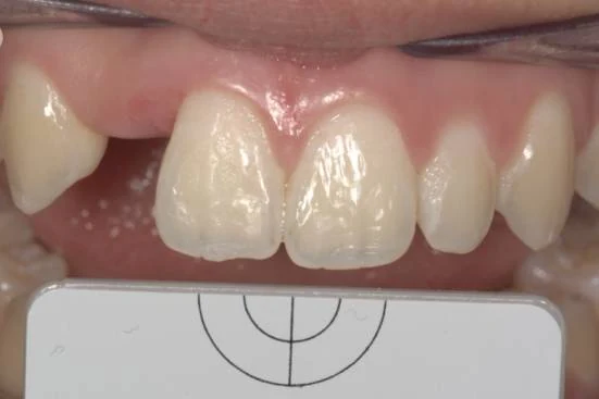 Before
 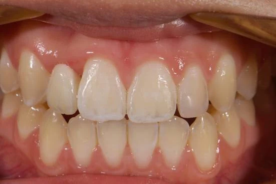 After
After Ceramic veneers, upper anterior segment. Shade taken from photographs rather than a tab, and the incisal translucency layered rather than stained on.
Ceramics Digital Dental Aesthetics Clinical work the referring practice
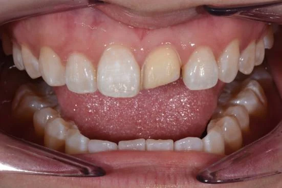 Before
 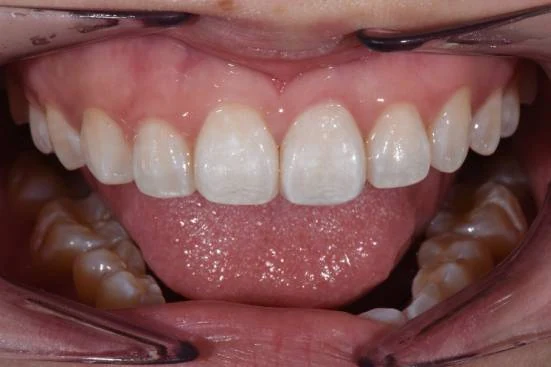 After
After Length and contour rebuilt across the upper anteriors. The line angles carry the light — that is what stops ceramic reading as flat.
Ceramics Digital Dental Aesthetics Clinical work the referring practice
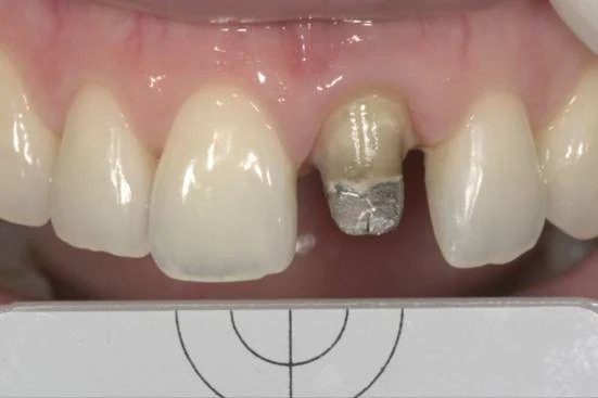 Before
 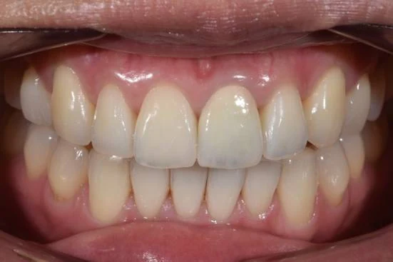 After
After A single central, matched to its living neighbour. The hardest case in ceramics, because there is nothing to hide behind.
Ceramics Digital Dental Aesthetics Clinical work the referring practice
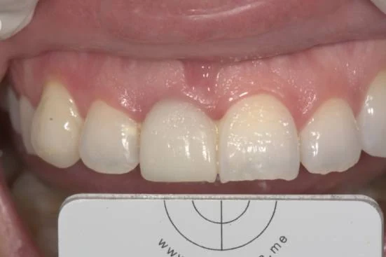 Before
 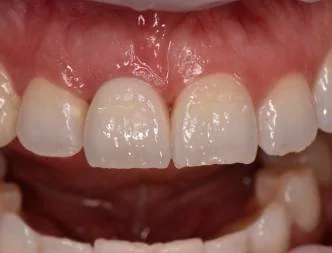 After
After Layered ceramic across the upper anteriors, built to a shape agreed at wax-up before anything was fired.
Ceramics Digital Dental Aesthetics Clinical work the referring practice
-
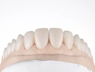
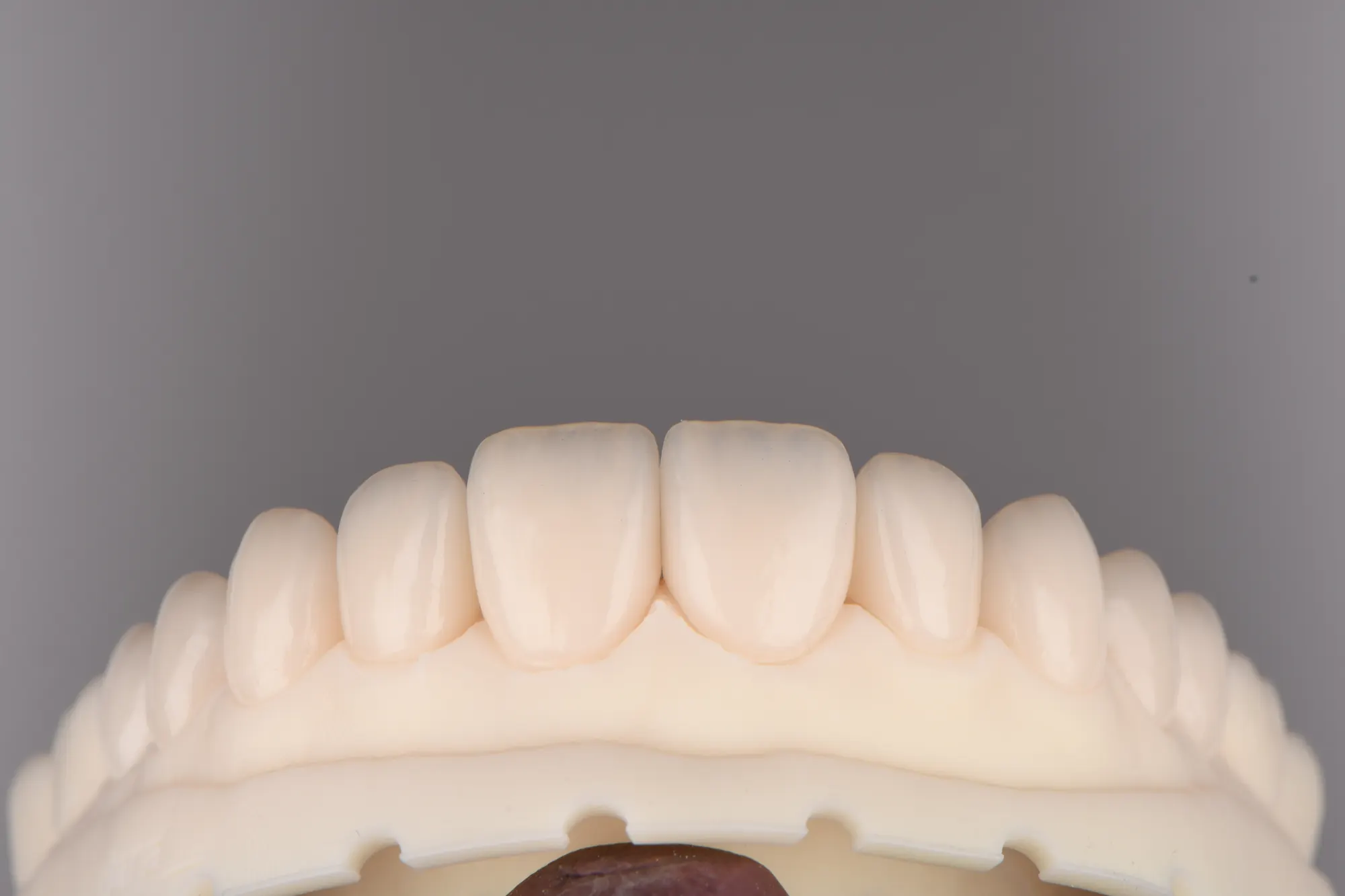
Ceramic veneers on the working model, ready for try-in. -
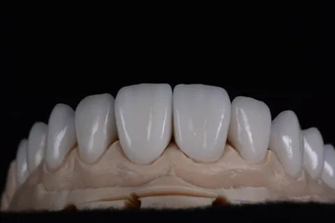
Layered anterior crowns — incisal translucency built in layers, not stained on. -
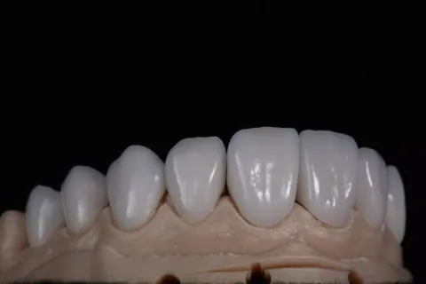
Anterior restorations checked on the model before dispatch. -
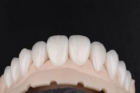
Anterior units, seated and margin-checked. -
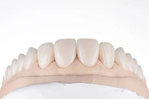
Shade assessment under controlled light before the case leaves the bench. -
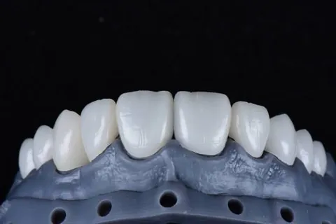
Anterior bridge — pontic contour worked to the ridge. -
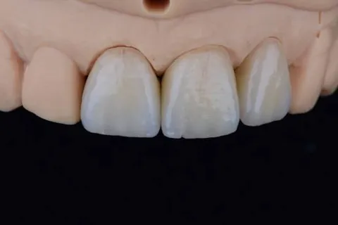
The hardest case in ceramics: one central, matched to its neighbour. -
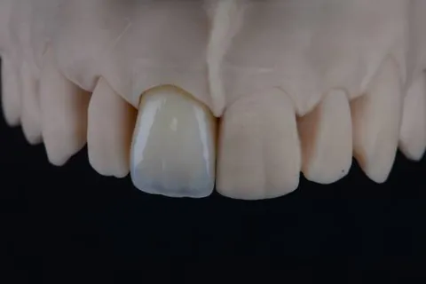
Try-in stage. Everything is still adjustable here — that is the point of it. -
Full-arch reconstruction, built to a planned occlusal scheme. -
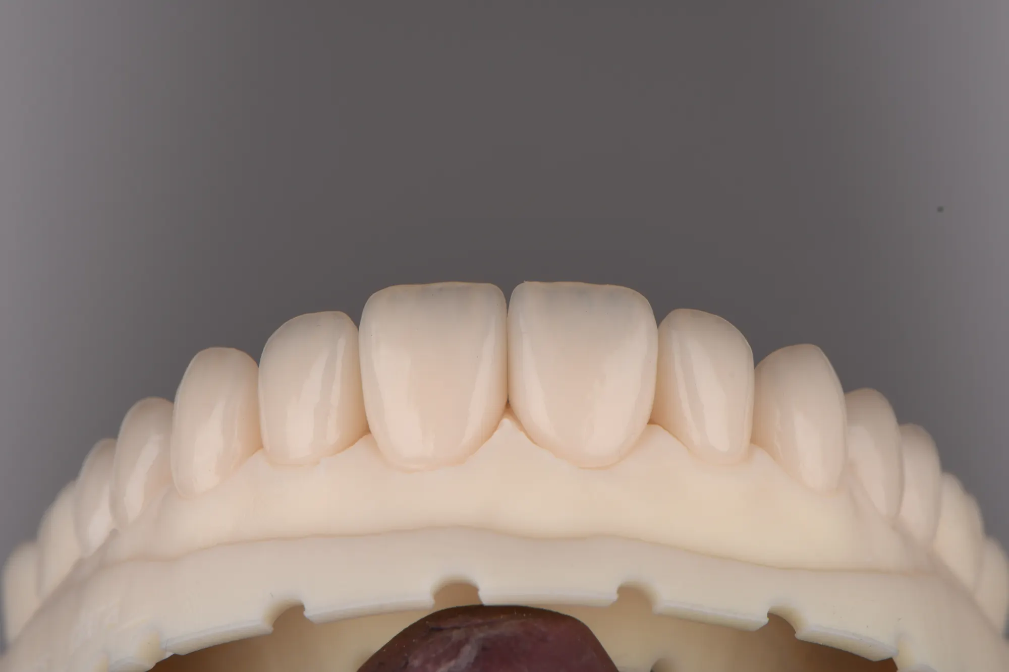
Surface texture and line angles — what makes ceramic read as enamel. -
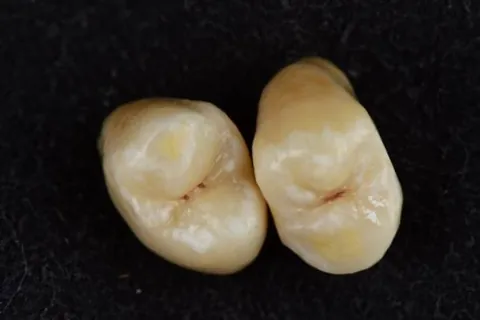
Posterior crowns — anatomy cut back and rebuilt for function first. -
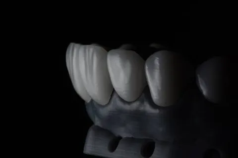
Zirconia bridge, checked on the die before glaze. -
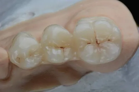
Occlusal detail — fissure pattern cut, not pressed flat. -
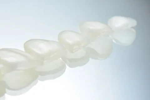
Monolithic posterior bridge where strength has to lead. -
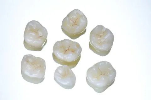
A set finished together, so they read as one mouth. -
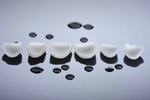
A day at the bench. -
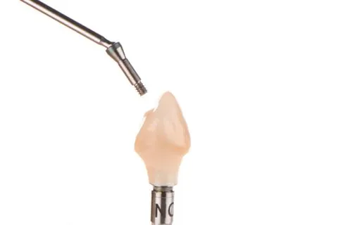
Implant crown on the holder, emergence profile shaped to the tissue. -
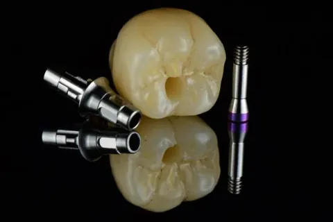
Crowns beside their abutments — the fit is decided here, not at the chair. -
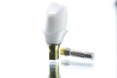
Seated on the abutment for verification. -
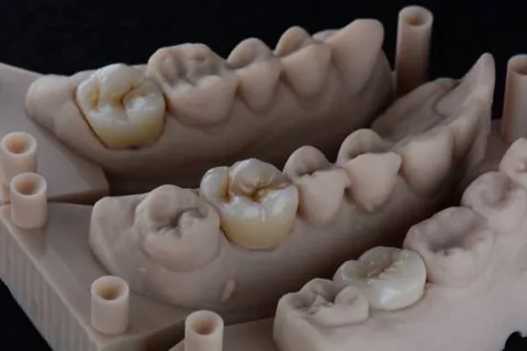
Articulated model — the case as the mouth actually closes. -
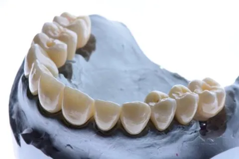
Full-arch working model with removable dies. -
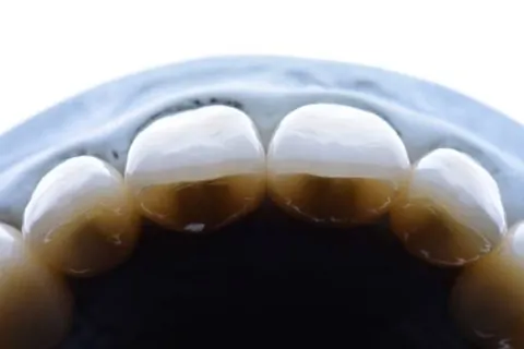
Margins. A lab is judged here before it is judged anywhere else. -
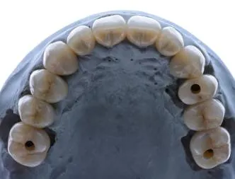
Occlusal view of the planned arch. -
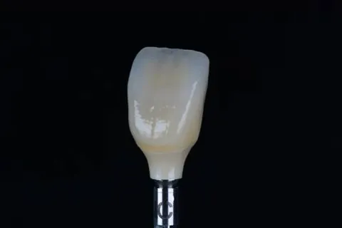
One crown, photographed the way we photograph every crown. -
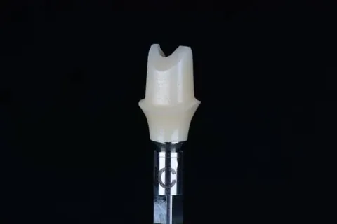
The inside of the crown — the surface nobody sees and everybody feels. -
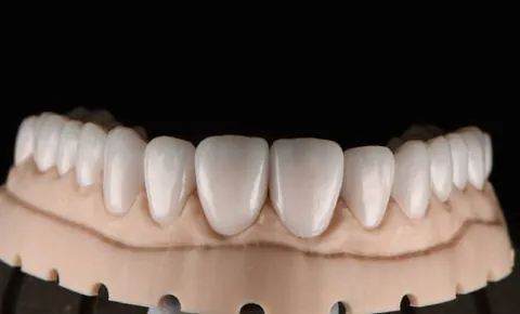
Digital wax-up — the shape agreed before a gram of ceramic is fired. -
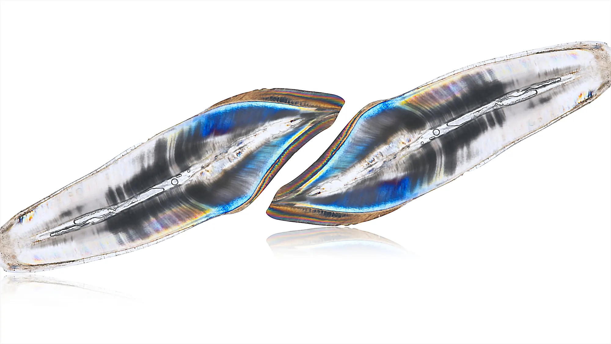
Veneer shells at 0.3mm. Held to the light, ceramic behaves like enamel — this is what shade tabs cannot show you. -
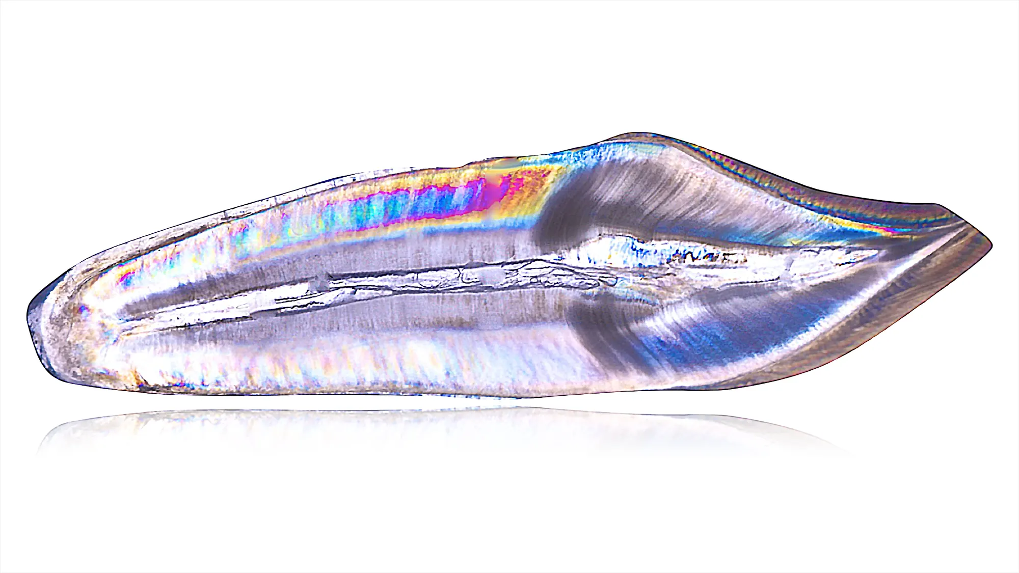
Opalescence in a single shell. The colour is in the material, not on it.
Nothing in that category yet.